
USD engineering faculty and students are using AI to improve cancer diagnostics
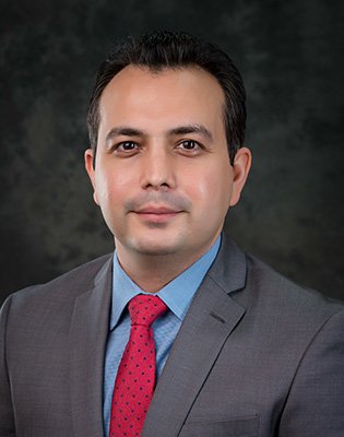
Meet Ebrahim Tarshizi, PhD, MBA, who is a Professor of Practice in the Shiley-Marcos School of Engineering and currently serving as Program Director for USD’s Applied Data Science (ADS) and Applied Artificial Intelligence (AAI) programs and as Director of the Center for Digital Civil Society (CDCS).
Prof. Tarshizi recently published a paper in the Journal of Pathology Informatics in collaboration with two graduate students in his MS-AAI program, Ken Devoe and Gary Takahashi, and a clinical pathologist, Alan Sacker, from Providence St. Vincent Medical Center in Portland, OR
Let’s take a few minutes to learn more about Prof. Tarshizi and this fascinating research.
How would you describe your recently published research to a 5 year old?
Imagine that you are a doctor who wants to help sick patients get better. There is a big machine that helps the doctor find out what's wrong with the patient by looking at pictures of where they are sick. But someone created a smaller machine, just like an iPhone, that might be able to do the same thing. Wouldn't it be great if that smaller machine could do the same thing? It would be easier to use, and maybe the doctor would use it more often than the larger machine. That's what we wanted to find out, and so we tested the smaller machine and found that it did a great job.
How did you get interested in this topic?
This topic was really led by a graduate student in one of our AAI classes, Gary Takashi. One of the topics we suggested for the final project in the AAI class on machine learning was exploring an image classification application in the medical world. Takahashi had experience with the MobileNet V2 model to identify patient pathologies, and wanted to see how well the more recent and improved MobileNet V3 architecture could be trained on pathology images of breast cancer. He also wanted to determine if it could distinguish between benign and malignant conditions, and classify their subtypes. For a doctor in practice who has little direct exposure to AI, this ability would seem like magic.
What's one challenge you faced in this research? How did you overcome it?
Machine learning models such as MobileNet V3 need to be trained on a set of images to accurately classify the image set of interest. But, training these models ‘from scratch’ would take a prohibitively long time to achieve accurate image classification. So, many of these models come already pretrained on ImageNet, a large database of images of everyday objects. However, the pretrained MobileNetV3 did not perform well on our pathology images. It was clear that we had to fine-tune the model on our own images, using a process called transfer learning, where we preserve some of the pretrained settings and fine-tune the rest. When we tested our fine-tuned model on the images in the dataset, the performance had improved substantially. The challenge was then to find the optimal balance of retaining pre-trained information and fine-tuning on our own pathology images, which required much trial-and-error. To address this challenge, Devoe developed a handy automated system of entering test settings in a spreadsheet, so that we could trial a variety of settings overnight, greatly accelerating the process. Then, I assisted the team with advanced data analytics to provide the necessary performance parameters.
What did you enjoy most about working on this project? Why?
While there have been publications describing the application of convolutional neural networks in the clinical pathology setting, most relied on intensive pre-processing to get accurate results. Other reports made use of larger network architectures, which require more hardware resources than the agile MobileNet series. Our report was one of the few that attempted to classify the subtypes of benign and malignant breast diseases, and it was exciting to see that this smaller model could perform excellent image classification on unenhanced pathology images.
What impact do you envision this work will have on society?
For one, I hope it will encourage other students interested in AAI to explore similar projects and ideas that have practical applications. Working with larger, proprietary image datasets, using an approach such as the one we described, it should be possible to develop a commercial model that clinical pathologists could use to identify a variety of diseases, much as computer vision is already helping diagnostic radiologists in reading digital mammograms.
What are you working on now?
Presently, I'm working on two new research projects and papers with my former students at USD. First, we are working on a machine learning project focused on detecting Alzheimer's disease using brain scan images. By employing neural networks, we aim to accurately classify and identify patterns in these images that are indicative of Alzheimer's, with the goal of improving early diagnosis and understanding of the disease's progression.
In the second project, we are addressing the prediction of hypotension in Intensive Care Units (ICUs), which is a critical indicator of patient morbidity and mortality. In this study, we are developing a deep learning model that utilizes high-resolution, minute-by-minute vital signs data to predict hypotension onset in ICU patients. This approach represents a significant advancement in early detection and intervention strategies for hypotension management.
We look forward to hearing more about these exciting projects!